Lawns of Flower Conservatory
“It is a beauteous evening, calm and free”
The Fracture of An Illusion
EXISTENTIAL ANGST
Selfish gene
A little girl's Art
^ ^
Religion explained
Farfalla
Two Tire economy
Art every where
Chopped !
An eye of a tree
Woods
Winter Sunrise
A page -- Nicholas Humphrey's "Soul Dust"
Mummy's pet, is that you....?
The Gregundrum / Figure 2
Time Flow
At Lands' End
Sylva
A photo session by Water Temple
A view across from Water Temple
Just watch -- I'll move it
Sulphrous landscape
Figure 8.4. T-O map, Leipzig, Eleventh century
Figure 12.6 Albert Einstein with Kurt Godel
Thrasymachus's challenge
Ockham’s Razor
From Bowels of Earth
Fun in the sun
Gotcha
Thinking
Mach's Ego Inspecting itself.
Melting glacier
One of the Peaks
A view from the Visitors center
Source of Sacramento river
Environmentally conscious...!
Wall of Sulphur
Keywords
Authorizations, license
-
Visible by: Everyone -
Attribution + non Commercial
- Photo replaced on 20 Oct 2019
-
9 visits
- Keyboard shortcuts:
Jump to top
RSS feed- Latest comments - Subscribe to the comment feeds of this photo
- ipernity © 2007-2024
- Help & Contact
|
Club news
|
About ipernity
|
History |
ipernity Club & Prices |
Guide of good conduct
Donate | Group guidelines | Privacy policy | Terms of use | Statutes | In memoria -
Facebook
Twitter


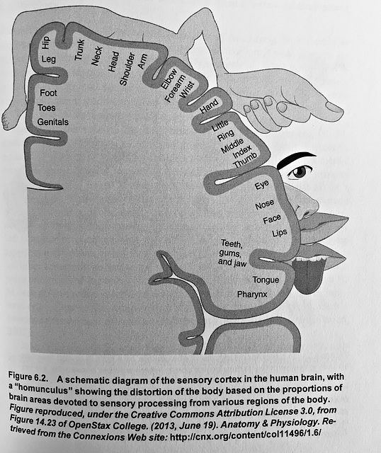
Control of movement is carried out by many regions of the brain, including some of the most evolutionarily primitive, such as the cerebellum and spinal cord, as well as some of the most evolutionarily recent. Such as the frontal cortex. Most sensorimotor processing (i.e., that which involves both sensation and movement) passes through an area of the neocortex (the evolutionarily recent top six neural layers of the brain). This area modulates perception and the processing of sensory stimuli (in the sensory cortex) and movement (in the motor cortex), and is collectively known as the sensorimortor cortex (Figure 6.2) Simulation of one area of the sensory cortex led a subject to experience a sensation in their pinkie finger, stimulation of an immediately adjacent area led to sensation in their ring finger and so on. These findings led to a representation of the human body’s topography with brain known as the “Penfield homunculus” -- a rather horrifyingly distorted cartoon with immense hands. The distortion arises because larger areas of the brain are dedicated to receiving inputs from the sending output to, more sensitive areas of the body (e.g., the fingers, tongue, genitalia, etc.).
The cortical homunculus is now known to be significantly more complex than Penfield originally envisioned, but it remains a cornerstone of both motor and sensory neuroscience (Schott, 1993). From the motor perspective, a revealing symptom of some epileptic seizures is the so called Jacksonian march, in which tremors move through the limbs in the precise order in which those limbs are encoded in the motor cortex; these areas are sequentially activated as the seizure activity moves through the brain. Neurosurgeons use a related concept during intraoperative testing, delivering weak electrical stimuli to areas of the motor cortex in order to precisely localize specific functions prior to surgery. ` Page 140/142
__________________________________________________________________________
The visual system is not the only place where the brain employs maps to organize spatial information. Much the same happens for your sense of touch. . . . . Each touch receptor monitors a particular region of the body surface, which is its receptive field. The signals from these touch receptors are sent along even longer axons, extending all the way from the receptor in the skin to the spinal cord. So individual neurons can be several feet long, from, say, the tip of your toe all the way to your spinal cord in your back! The axons from the same location travel together and from synapses of neighboring neurons in the spinal cord, which in turn send axons to neighboring regions in the body region of the thamalus and from thence to the cortex. All along the way, the pattern of input continues to match the pattern of locations of the body surface, so the neurons that receive input originating in the toe, elbow, or nose are clustered together in distinct zones. The body surface maps of your skin is thus duplicated at each stage along the neural road into the brain. In the ‘somatosensory’ cortex, the cortex responsible for body-related information, . . . . Page 87
Penfield stimulated all over the brain in his patients, and he asked them to describe what they noticed. Patients reported a variety of sensations -- sights, sounds, touch -- depending on where the electrode was placed. When Penfield stimulated the somatosensory cortex, the sensation would felt at a particular location of the body surface. Moving the electrode to a slightly different location would move the sensation to an adjacent location on the body. Penfield thus built up detailed maps of how the body surface was represented to the somatosensory and nearby motor cortex that are still in use today. ~ Page 104
Sign-in to write a comment.